
Hotline:
400-863-1908Monday to Friday: 9:00-18:00
Traumatic Brain Injury (TBI) refers to damage to the brain caused by external mechanical forces, with its clinical incidence increasing year by year. To better study the pathogenesis of these issues, understand the pathophysiological process of craniocerebral trauma, and develop new treatment methods, establishing a high-quality animal model is of crucial importance.
A self-modified free-fall method was used to establish a severe craniocerebral injury model in rats.
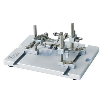
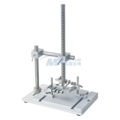
Free-fall method: It consists of a stereotaxic instrument, a fixing frame, a smooth stainless steel catheter with holes, a falling weight, an impact needle, and a baffle. During use, the baffle is inserted into the stainless steel catheter at a certain height, the weight is placed in the catheter, and the impact needle is placed on the dura mater. The baffle is quickly pulled out, and the weight impacts the impact head at a free-fall speed. The impact head then directly strikes the dura mater to establish a craniocerebral injury model. The following are some accessories of the free-fall impactor.
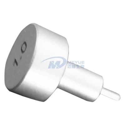
Impact Needle (Select the corresponding model of impact needle according to the size of the rat)

Weight (Select the weight corresponding to the size of the rat)
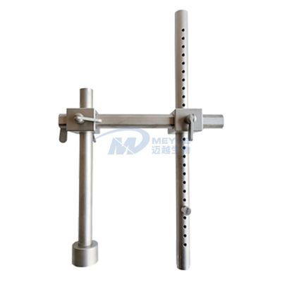
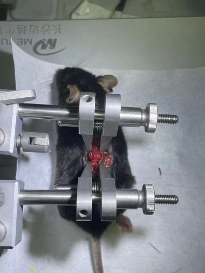
Free-Fall Bracket (Optional accessory)
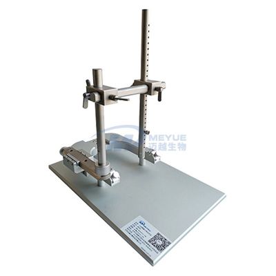
Spinal Impact Adapter (Used to fix the rat's spinal cord)
The following is the operational procedure
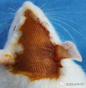
Male Sprague-Dawley (SD) rats weighing 250-300g were selected, and fasted for 8-12 hours before modeling. The rats were weighed and anesthetized with 10% chloral hydrate (0.3ml/100g); alternatively, if conditions permit, inhalation anesthesia with gas anesthetics can be used to reduce the mortality rate of rats while ensuring the success rate of the experiment. A hair clipper was used to shave the hair on the top of the rat's head for skin preparation. After routine disinfection with povidone-iodine, a midline sagittal incision was made on the scalp, with a length of approximately 3-4cm.
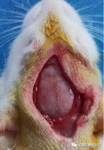
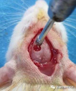
The soft tissues were dissected, the periosteum was stripped off, and the left parietal bone was exposed. A small hole was drilled with a high-speed skull drill at 3mm posterior to the coronal suture and 2.5mm left lateral to the midline (this position can be adjusted), which was then expanded into a bone window with a diameter of 5mm. The arachnoid membrane was exposed while keeping the dura mater intact.
The impact needle was placed outside the dura mater, with no weight added at this time. The impact needle was adjusted upward by 2-3mm, so that when a 40g weight was dropped freely along the catheter from a height of 20cm to impact the impact needle (by suddenly pulling out the baffle), the compression depth would be 2-3mm without penetrating the dura mater, resulting in local cerebral contusion. The impact force was 800g·cm (40g × 20cm). After the impact, the impactor was immediately removed from the injury site to avoid secondary injury. The scalp was sutured layer by layer, the sutured wound was clamped with hemostats, wiped with alcohol cotton balls, and the ear bars and the rat were removed. Penicillin was injected intramuscularly for anti-infection. The animal was placed back into the rearing cage, and attention was paid to keeping it warm after the operation.
Previous: Technology of Stereotaxic Instrument and Specific Operation Method for Rat and Mouse Experiments
Next: Experimental operation method of micro drug delivery cannula
Copyright © 2019 Changsha Meyue BlO co., Ltd Hunan ICP Filing 19018469-1
Address:No. 186 Guyuan Road, Hunan University Science and Technology Park, High tech Zone, Changsha City Hotline:400-863-1908 Consultation:19892896528(Zhang)、19892863908(Hu)、19918939165(Jiang) Email:my@meyuecn.com

Follow Us
 简体中文
简体中文
 English