简体中文
简体中文
 English

Electron microscopes, with their high resolution, can clearly present the fine structures of the microscopic world, enabling researchers to conduct in-depth studies at the cellular and molecular levels. They provide key data and insights for scientific research in multiple fields such as biomedicine and materials science, making them crucial in the scientific research industry.
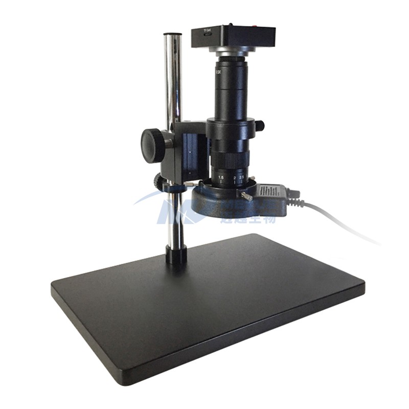
Meyue Bio M5274 Electron Microscope is equipped with a Sony high-pixel camera, featuring 2K resolution and 2560×1440 shooting pixels. It comes with an HDMI interface, a parfocal zoom lens, and a metal bracket, boasting rich functions. It provides accurate microscopic observation for scientific research and other fields, meeting your scientific research needs.
Performance Features
| Feature Category | Detailed Description |
|---|---|
| Camera Configuration | Equipped with a Sony 1/3' 2μm x 2μm camera, which ensures image clarity and detail capture capability through high-pixel performance. |
| Imaging Parameters | 2K high-definition resolution with a shooting pixel of 2560×1440, capable of presenting accurate and detailed microscopic images, meeting the strict requirements of scientific research for high-resolution images. |
| Interface Design | Adopts an HDMI high-definition output interface, which can be easily connected to a display to realize high-definition display of microscope images, facilitating observation and analysis. |
| Lens Characteristics | Equipped with a 0.7X~4.5X optical parfocal lens, enabling 20~180x parfocal continuous zoom. The parfocal function ensures no need to refocus when switching magnifications, greatly improving observation efficiency. |
| Bracket Structure | A lifting bracket made of 10A metal material, with excellent stability and durability, providing reliable support for the stable operation of the microscope. |
| Software System | Built-in Linux measurement system. |
| Equipment Accessories | Including an LED high-brightness fill light (brightness can be adjusted according to sample characteristics and observation environment to provide sufficient and uniform illumination) and an HDMI high-definition video cable. |
System Functions
| Function No. | Function Description |
|---|---|
| 1 | Supports Chinese and English switching. |
| 2 | Built-in measurement function, enabling accurate size measurement of microscopic structures. |
| 3 | Automatic edge-finding function can quickly determine the sample boundary, improving measurement efficiency. |
| 4 | Data export function facilitates the use of measurement data for in-depth analysis later. |
| 5 | Freeze-frame function allows researchers to carefully observe images at specific moments. |
| 6 | Split-screen comparison (2/4 split-screen) function can compare multiple samples or different areas of the same sample at the same time, helping to analyze differences. |
| 7 | Equipped with 12 groups of crosshairs and scales for sample positioning and size measurement. |
| 8 | Mirror function meets different observation habits. |
| 9 | Parameters such as white balance, exposure, color, contrast, and sharpness can be adjusted to optimize image quality according to sample characteristics. |
| 10 | Supports USB flash drive storage, enabling operations such as screenshot, photo shooting, and video recording, which is convenient for recording experimental data and microscopic phenomena. |